Treatment and Surgery Restoration of Urology Lithiases
Cystoscopy
Cystostopy is an endoscopic technique which is considered one of the most important “diagnostic urological tests”, for the examination of the lower urinary system. The test takes a short time, is carried out with local anaesthesia and can be performed in the urologist’s private office, provided it is equipped with the proper endoscopic equipment. The test uses a flexible cystoscope with a diameter of a few millimeters, and can check the urethra for damage in the sphincter or narrowing, the urinary bladder walls, the ureter mouths, the prostate size in men, or whether there are stones, a tumor or growth in the urinary bladder. During the cystoscopy, tissue samples can be collected for biopsy, and some pathological damages can be cauterized. After cystoscopy, the patient may experience mild nuisance (burning sensation) during urination, as well as frequent urge to urinate. These problems do not last more than 2-4 hours and are treated with common pain killers. video
Urethroscopy
Urethroscopy is an endoscopic technique which is considered one of the most important “diagnostic urological tests”. The test takes a short time, is carried out with local anaesthesia and can be performed in the urologist’s private office, provided it is equipped with the proper endoscopic equipment. The test uses a flexible cystoscope with a diameter of a few millimeters, and can check the urethra for damage in the sphincter or narrowing, inflammation or exophytic lesion. During the urethroscopy, tissue samples can be collected for biopsy, and some pathological damages can be cauterized. After urethroscopy, the patient may experience mild nuisance (burning sensation) during urination, as well as frequent urge to urinate. These problems do not last more than 2-4 hours and are treated with common painkillers. video
PIGTAIL Catheters (Ureteral Self-Supporting Catheters) Placement
The PIGTAILS or ureteral self-supporting catheters are named after the size of their tightly curled ends, which resemble a pig’s tail. They are used in order to keep open the urine flow channel which has been blocked by stones or newly formed growths of the upper and middle urinary system, stenoses or injuries of the ureter etc., which result in increased morbidity (renal colic, prolonged and/or accompanied by fever, and renal failure). Their placement is usually performed in surgery with X-ray test in aseptic conditions. After the operation the patient must remain in hospital for a few hours for observation.
Ureter-Kidney Stone – Endoscopic Removal
Nowadays, the treatment of upper and middle urinary system lithiases demands minimally invasive approach. The second most common intervention is the endoscopic removal of a ureter-kidney stone. With a special instrument, the ureteroscope, the stone that causes the blockage is identified and with a special technique and proper handling is immediately crushed with the use of ultrasounds, electrohydraulic waves or laser, depending on the position, shape and composition of the stone. The operation is carried out with light sedation or epidural anaesthesia and its duration depends on the size, position and composition of the stone. In some cases it is advisable that a ureteral self-supporting catheter (PIGTAIL) be placed. The patient must remain one day in hospital for observation, while morbidity of the operation is minimal.
Ureter-Kidney Stone – Extracorporeal Shock Wave Lithotripsy
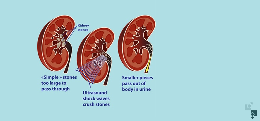
The Extracorporeal Shock Wave Lithotripsy (ESWL) is a non-invasive approach to the treatment of the upper and middle urinary system. The extracorporeal lithotripter is a high-technology instrument for the crushing of calculi by the use of shock waves, and is considered the safest treatment method. The entire procedure is carried out with the use of mild analgesics, the patient can immediately return to their normal activities, while it is possible that the procedure may need to be repeated within 20 days. video
Kidney Stone - Percutaneous Nephrolithotomy
Nowadays, the treatment of upper and middle urinary system lithiases demands minimally invasive approach. A modern way for the removal of a kidney stone is the Percutaneous Nephrolithotomy (PCNL). A percutaneous paracentecis of the kidney is performed under X-ray guidance. Next, a special instrument, the nephroscope, is incerted and identifies the stone that causes the blockage. Then, with a special technique and the proper handling, the stone is immediately crushed with the use of ultrasounds, electrohydraulic waves or laser, depending on the position, shape and composition of the stone. The operation is carried out with light sedation or epidural anaesthesia and its duration depends on the size, position and composition of the stone. In some cases it is advisable that a ureteral self-supporting catheter (PIGTAIL) be placed. The patient must remain one day in hospital for observation, while morbidity of the operation is minimal.
Ureter-Kidney Stone – Open Surgery
The open surgery for the treatment of kidney and ureter stones has been significantly limited to cases of coral-like stones or failure to face the problem with the modern methods. The operation is carried out with general anaesthesia. The area of the lithiasis is approached surgically and a ureterolithotomy – pelvolithotomy – nephrolithotomy (depending on the stone’s position) is performed. The stone is removed and then a ureteral self-supporting catheter (PIGTAIL) is placed. In case there co-exists an anatomical damage, it is treated surgically. The surgical trauma is stitched and drainage is placed for 1-2 days. The patient must remain in hospital for observation for 2-4 days, while morbidity is minimal.
Ureter-Kidney Stone – Laparoscopic Removal
The laparoscopic removal of kidney and ureter stones is a modern technique for the treatment of such conditions. Η λαπαροσκοπική αφαίρεση λίθων είναι μια μοντέρνα μέθοδος αντιμετώπισης του νεφρού και του ουρητήρα έχει περιοριστεί σημαντικά και διενεργείται μόνο σε περιπτώσεις κοραλιωειδών λίθων ή αδυναμία - αποτυχία επίλυσης του προβλήματος με τις σύγχρονες μεθόδους αντιμετώπισης. The procedure is carried out with general anaesthesia. Without need for opening the abdominal walls, the surgeon reaches the area of lithiasis and performs a ureterolithotomy – pelvolithotomy – nephrolithotomy (depending on the stone’s position) is performed. The stone is removed and then a ureteral self-supporting catheter (PIGTAIL) is placed. In case there co-exists an anatomical damage, it is treated surgically. The surgical trauma is stitched and drainage is placed for 1-2 days. The patient must remain in hospital for observation for 2-3 days, while morbidity is minimal.
Urinary Bladder Stone – Open Surgery
The open surgery for the treatment of urinary bladder stones has been significantly limited to cases of coral-like stones or failure to face the problem with the modern methods. The operation is carried out with general anaesthesia. The area of the lithiasis is approached surgically and a cystolithotomy is performed. The stone is removed and then a urinary bladder catheter is placed. In case there co-exists a benign prostatic hyperplasia, the prostatic gland is removed surgically. The surgical trauma is stitched and drainage is placed for 1-2 days. The patient must remain in hospital for observation for 2-4 days, while morbidity is minimal.
Urinary Bladder Stone – Endoscopic Removal
Nowadays, the treatment of urinary bladder lithiases demands minimally invasive approach. The most common intervention is the endoscopic removal of a urinary bladder stone. With a special instrument, the cystoscope, the stone that causes the blockage is identified and with a special technique and proper handling is immediately crushed with the use of ultrasounds, electrohydraulic waves or laser, depending on the position, shape and composition of the stone. The operation is carried out with light sedation or epidural anaesthesia and its duration depends on the size, position and composition of the stone. In some cases it is advisable that a urinary bladder catheter be placed. The patient must remain one day in hospital for observation, while morbidity of the operation is minimal.


